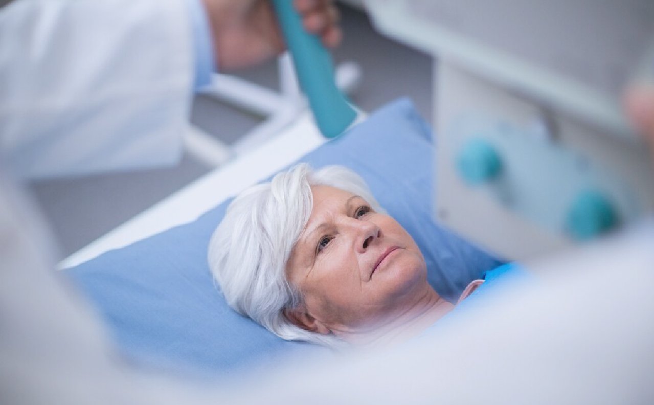
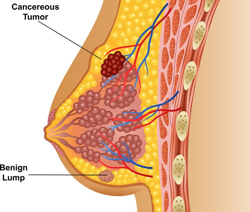
Breast cancer diagnosis often begins with an exam and a discussion of your symptoms. Imaging tests can look at the breast tissue for anything that's not typical. To confirm whether there is cancer or not, a sample of tissue is removed from the breast for testing.
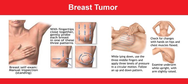
Breast exam: During a clinical breast examination, a trained healthcare provider carefully examines the breasts, observing for any abnormalities such as changes in skin texture or nipple appearance. They then use their fingers to palpate the breasts, checking for any unusual masses or lumps. Additionally, they perform a thorough examination of the collarbones and the areas surrounding the armpits to ensure comprehensive assessment for any concerning findings.
Mammogram:
A mammogram utilizes X-rays to capture images of breast tissue, commonly employed as a screening tool for detecting breast cancer. Should an initial screening mammogram reveal any abnormalities, a follow-up examination known as a diagnostic mammogram may be recommended. This in-depth procedure allows for a closer examination of both breasts to further investigate any suspicious findings.
Breast ultrasound:
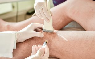
Ultrasound technology utilizes sound waves to generate images of internal body structures. When it comes to assessing a breast lump, a breast ultrasound provides valuable insights to healthcare professionals. It can distinguish between a solid mass and a fluid-filled cyst, aiding in the diagnostic process. This information guides healthcare teams in determining the subsequent tests that may be necessary for further evaluation.
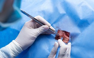
Removing a sample of breast cells for testing: A biopsy is a procedure to remove a sample of tissue for testing in a lab. To get the sample, a healthcare professional puts a needle through the skin and into the breast tissue. The health professional guides the needle using images created with X-rays, ultrasound or another type of imaging. Once the needle reaches the right place, the health professional uses the needle to draw out tissue from the breast. Often, a marker is placed in the spot where the tissue sample was removed. The small metal marker will show up on imaging tests. The marker helps your healthcare team monitor the area of concern.
Breast tumor removal by VATS is suitable for individuals with symptomatic thyroid nodules who are seeking an alternative to conventional surgical options. Consultation with an interventional radiologist can help determine if Breast tumor removal by VATS aligns with your health needs and medical history.
You should make an appointment by calling or by email
Make an Appointment +91 960 645 9782